INTO THE TISSUE

MRI powers studies of ACLs, Alzheimer’s and athletes – and unique educational opportunities


UNCG researchers are using a magnetic resonance imaging machine, or MRI, to peer inside joints and take pictures of study volunteers’ brains. Their goal is to better understand how our bodies and brains function – and why things go wrong – to find ways to prevent common problems.

“It never ceases to amaze me that here at UNCG we have access to this amazing tool to help us ask the questions that we really want to ask,” says Randy Schmitz, professor of kinesiology and director of musculoskeletal imaging at the Gateway UNCG MRI Center, where the MRI is housed.

“This has torn down barriers,” he says. “Our researchers no longer have to go to a medical institution here in town or another academic institution 60 miles away.”

MRIs create high-resolution images of tissues that have water in them – virtually all tissues in the human body.

Powerful magnets in the machine cause protons in water molecules to align, and then the machine emits radio waves that tickle those water molecules. The water molecules give off energy the machine detects to create pictures of tissues. It can even measure blood flow in the brain.

Tools of the Trade

What do 55-million-year-old fossils, shrews with shrinking skulls, studies on Alzheimer’s and exercise’s impacts on the human brain, and tiny tech for a less toxic world have in common? The cutting-edge new tools driving discovery at UNCG.

MRIs create high-resolution images of tissues that have water in them virtually all tissues in the human body.

Powerful magnets in the machine cause protons in water molecules to align, and then the machine emits radio waves that tickle those water molecules. The water molecules give off energy the machine detects to create pictures of tissues. It can even measure blood flow in the brain.

Schmitz and Dr. Sandy Shultz, another kinesiology faculty member, use the machine in their study of anterior cruciate ligaments, which connect the thigh bone to the shin bone. ACL injuries are common in the United States among both athletes and nonathletes.

They are interested in figuring out why some people might be more prone to ACL injuries. Differences in knee and ACL structure could be the key, and might lead to better methods of injury prevention.

Schmitz and Shultz with their patented device to test knee laxity

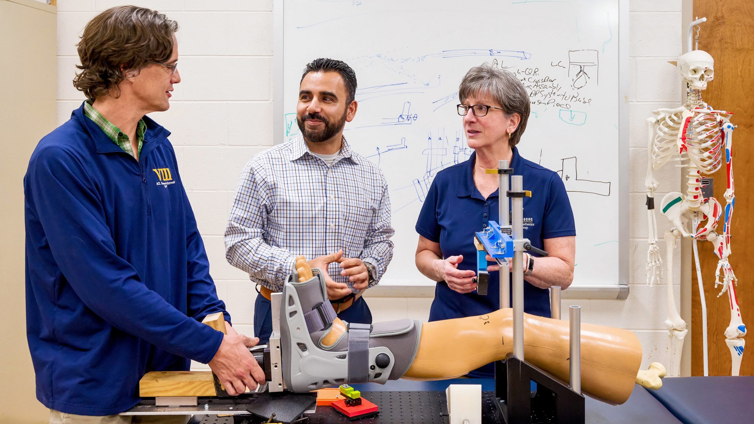
Recently Schmitz (l) and Shultz (r) patented a device to measure knee laxity – a strong predictor for future injury among young athletic females – across all three axes of motion.

Other researchers are using the MRI machine to investigate interactions between the brain and the body.

Dr. Jennifer Etnier, professor of kinesiology, is exploring how exercise may delay the onset of Alzheimer’s. In one large research study with $3.4 million in National Institutes of Health funding, she recruited middle-aged and older adults who may have a genetic risk of Alzheimer’s disease.

MRI scans of the volunteers’ brains are one of the ways Etnier is assessing the impact of exercise on the volunteers’ Alzheimer’s risk. Among people with the highest genetic predisposition for Alzheimer’s, brain changes can be detected as early as in someone’s 40s. Etnier hopes to be able to assess whether exercise delays those harmful changes in brain structure.

  • Alexis Ganesh and Etnier with UNCG's MRI machine
  • Researchers reading data on a computer
  • A patient stretching her arms using a green rubber band
  • Researchers looking at data on a laptop

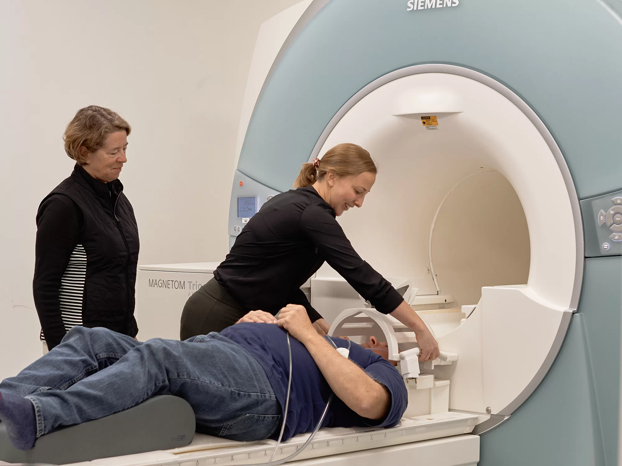
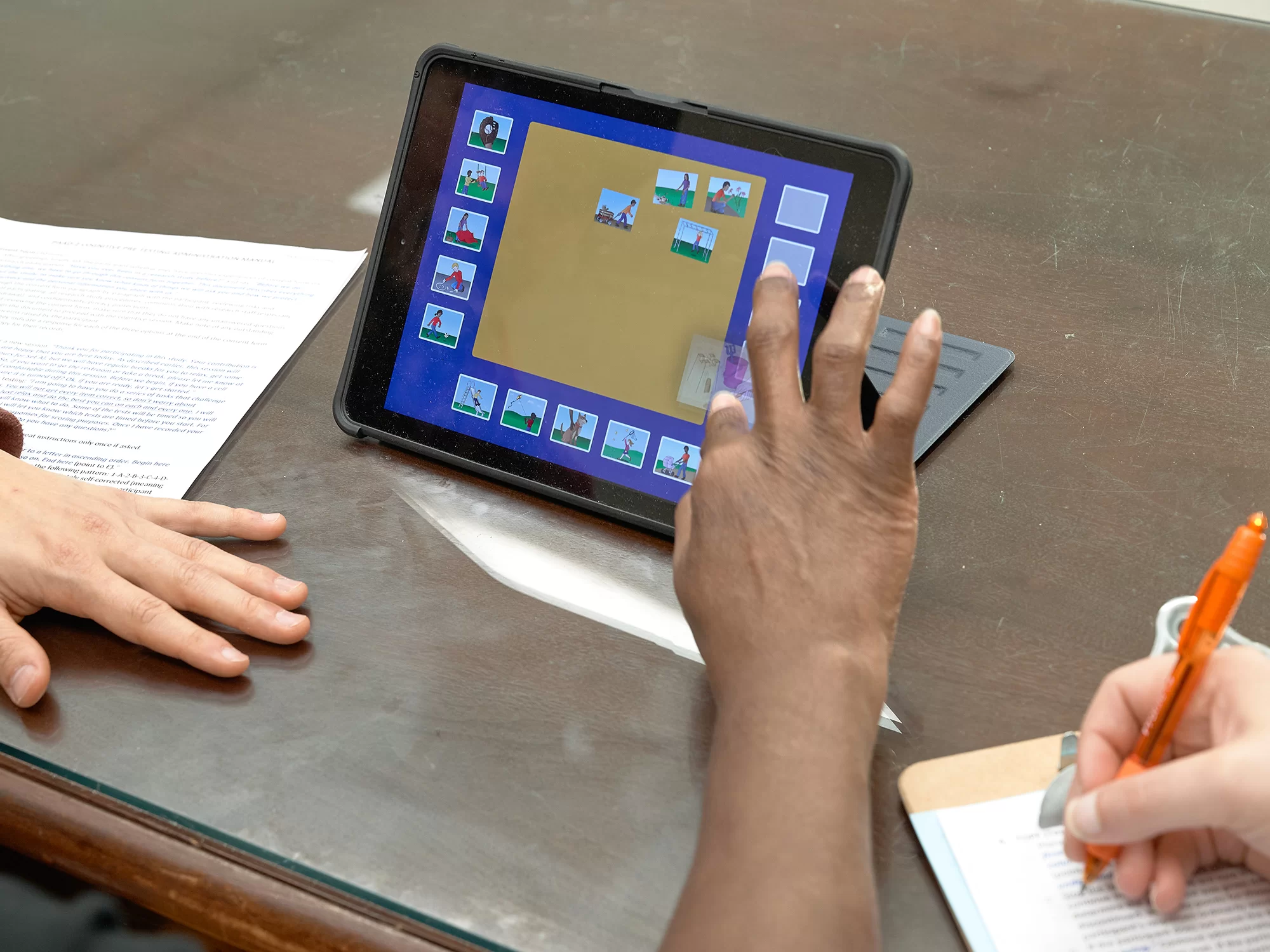
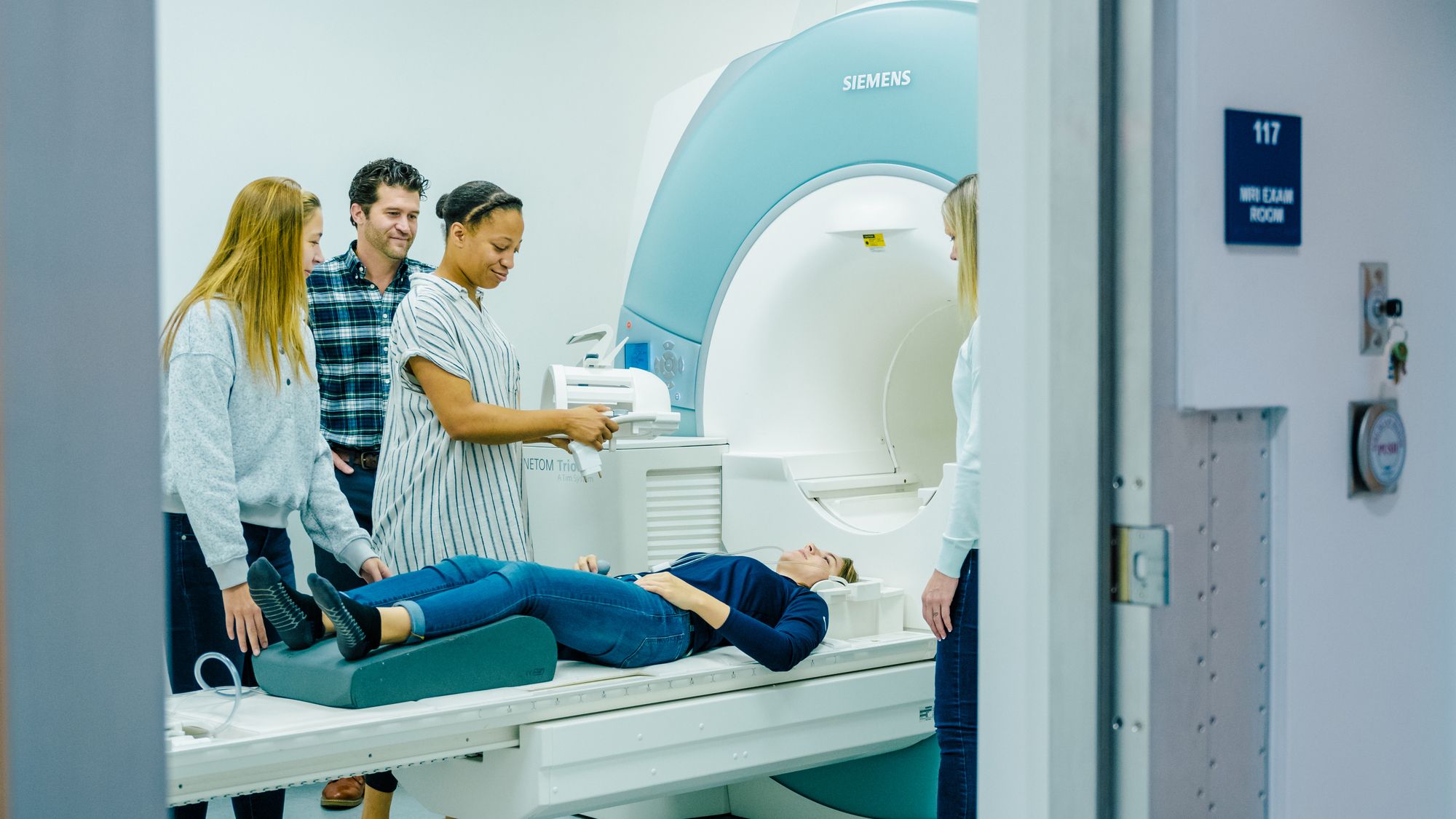
“From the first day of my doctoral program, I was excited to use the MRI scanner,” says Dr. Alexis Ganesh, who worked with Etnier (photo 1) and UNCG’s machine as a grad student and then as a postdoc.

Psychology faculty member Brittany Cassidy has used the MRI to study what parts of the brain are involved in different activities involving learning and memory.

One study, for example, examined how connectivity in the brain’s default network affects mnemonic discrimination — an important kind of memory we use in daily life.

“It is looking at whether you can distinguish existing memories from similar perceptual experiences,” Dr. Cassidy says. “For example, on a table full of coffee mugs, how can you remember which one is yours when you get back from a coffee break?”

Cassidy, along with her undergraduate and graduate researchers, has also used the MRI to study why older adults tend to be more trusting of others than younger adults — especially when the other person behaves negatively.

The MRI showed that during the learning process, different parts of older adults’ brains were more active when learning about someone’s reputation for trustworthiness, or lack thereof.

“These are opportunities that don’t happen at every university and a strength of UNCG,” says Cassidy. “Students get to really see what’s going on versus just learning about the methods in the classroom, and that can be really, really eye opening.” 

Brain-body connections are also the research focus of kinesiology’s Derek Monroe. He’s trying to better understand the physiological and neurological results of traumatic brain injuries

“After a head injury, you might assume that damage will be localized to brain areas near to where the impact occurred,” Dr. Monroe says. But his studies, conducted across different sports and populations, hint at more widespread effects.

“In my preliminary findings, the processes that regulate our basic bodily functions seem to be affected. However, there is still a lot of work to be done for us to understand the when, why, and how.”

Monroe and his collaborators combine MRI data with information from the field, such as symptom reports and impact data recorded by specialty mouthguards.

He’s also interested in the benefits of exercise. “We’re exploring how exercise can help brains recover from injury and how it contributes to general brain health across our lifespan.”

Monroe, whose focus is on behavioral neuroscience, was drawn to UNCG in 2020 after a postdoc at the University of California-Irvine medical school.

“The MRI, the facility, the resources, and other faculty who were already users of the technology are a big reason why I got excited about the position here at UNCG,” he says.


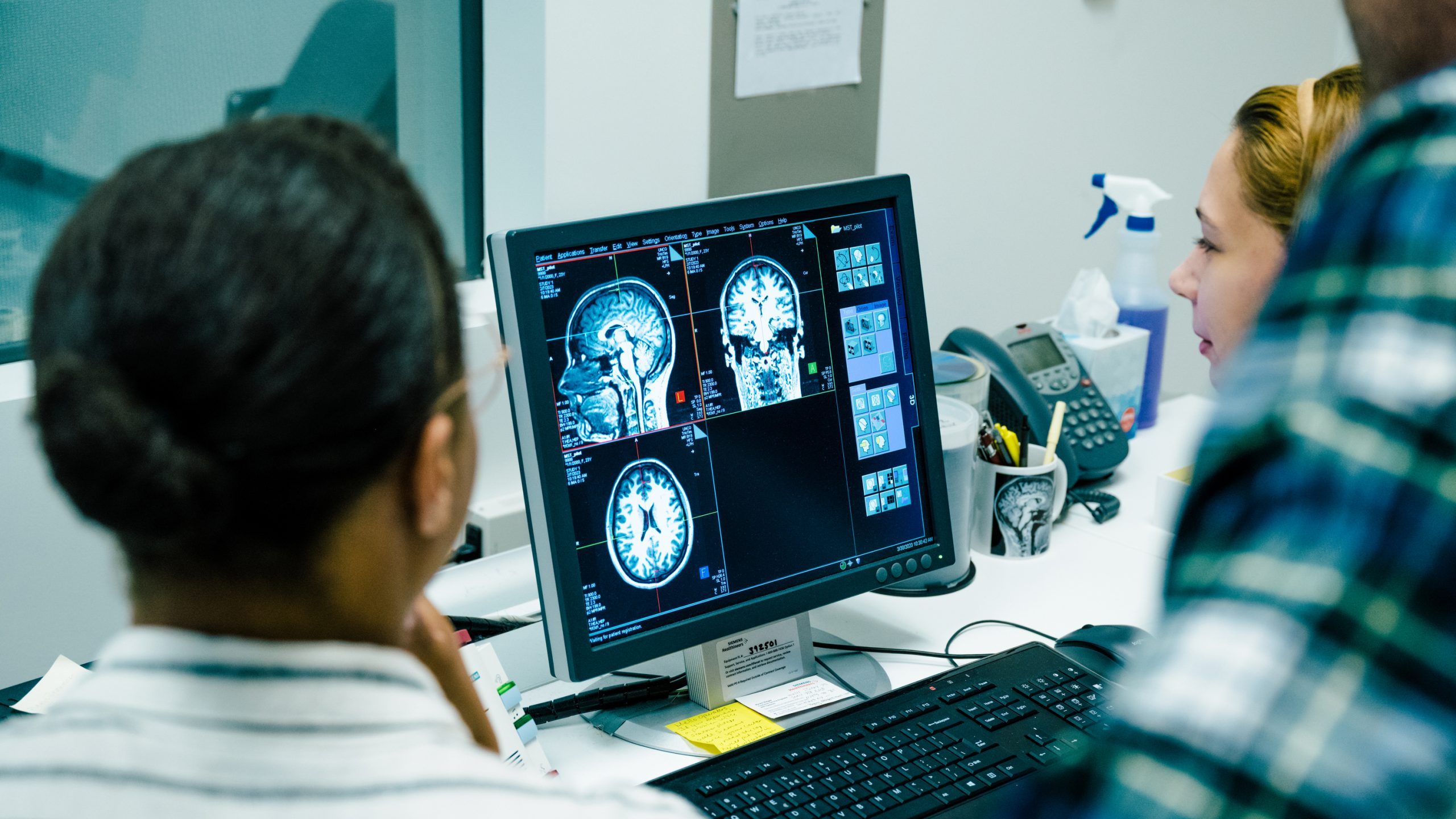
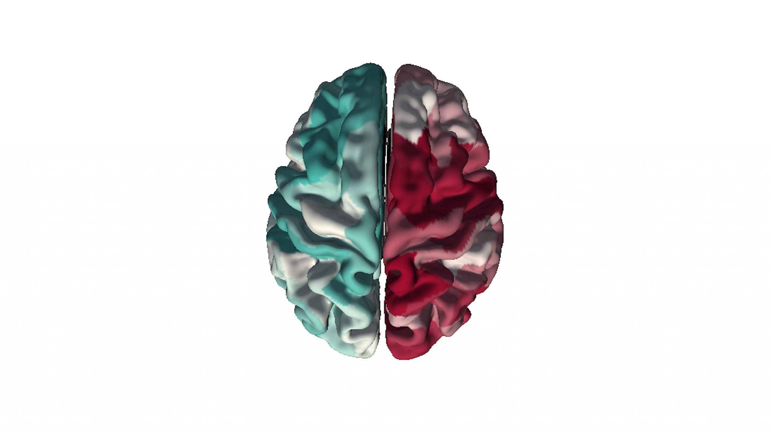
When examining athletes experiencing severe post-concussive symptoms, Monroe and his team found differences in the shapes of some parts of the brain. The structural changes the team found, seen below on the left, correlate with a pattern of “risky” gene expression previously identified in post-mortem studies of brains, which is visualized on the right. 

Top view of the human brain with one red and white side and another green and white
Front view of human brain with one side that is yellow and orange and the other multicolored

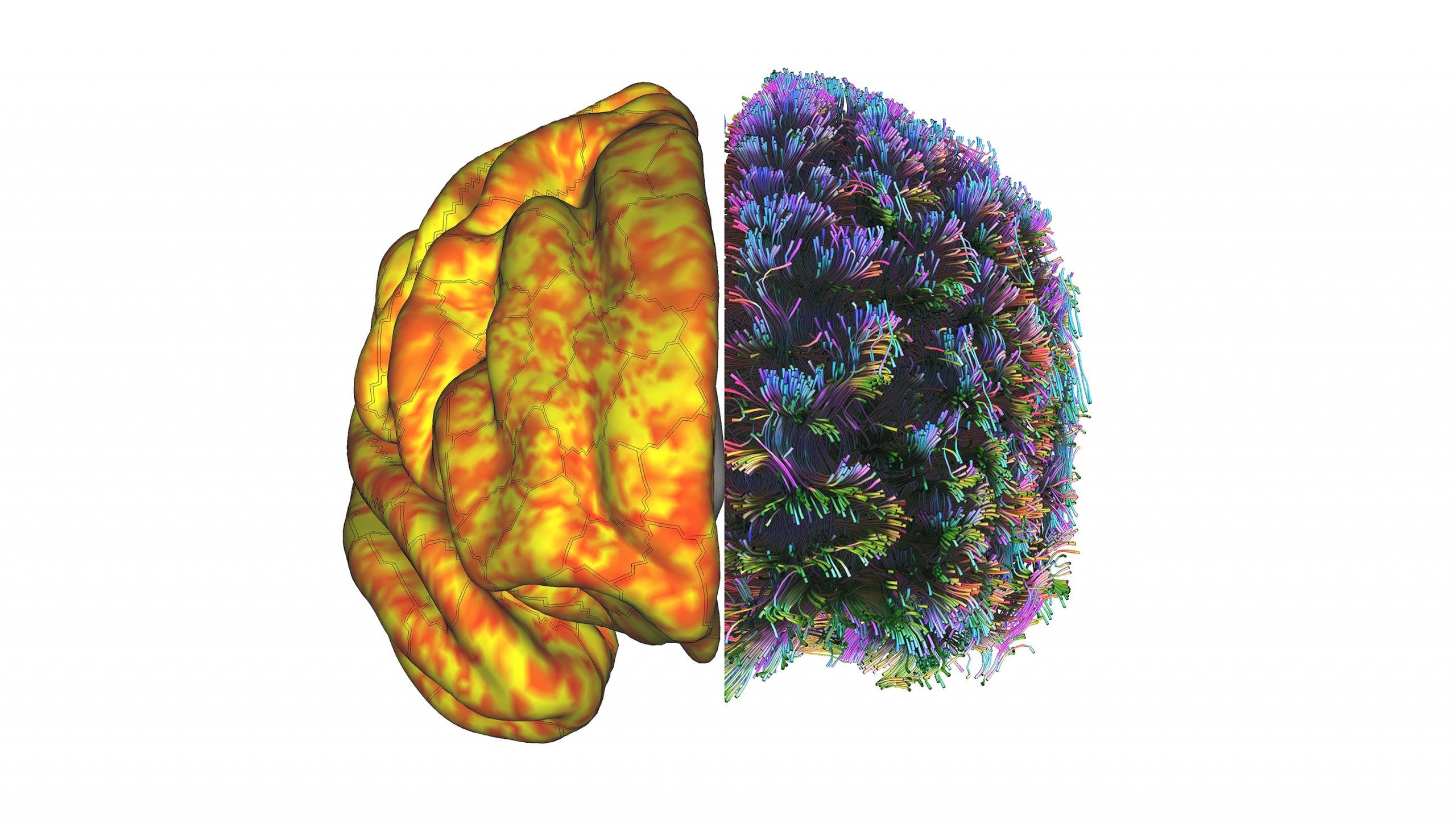
Above on the left is a map Monroe generated of the grey matter surface of a subject’s brain. Areas in orange are functionally connected while the subject is in a resting state. On the right is a diagram of how deeper, white matter fibers connect different parts of the brain. Monroe is interested in how exercise shapes brains. For example, Monroe and Dr. Donna Duffy in kinesiology found that roller derby athletes at rest exhibit unique brain patterns. 


Monroe is also teaching students how to use the MRI machine.

“I want to get graduate students now, and hopefully undergraduates in the near future, into courses – get them exposure to a machine that they would not have access to anywhere else,” he says. “In the graduate course we spend two weeks, and we go over and we measure our brains. We then spend the second half of the course analyzing our brains.”

That course involves more than just learning how to operate the machine. Students must learn about several disciplines, including physiology, physics, psychology, and data science. They learn how to upload the data and analyze it on powerful computers, and how to ask scientifically valid questions and understand the answers the MRI provides.

None of that, Monroe says, would be possible without both the equipment and a UNCG culture that encourages learning and interdisciplinary collaboration.

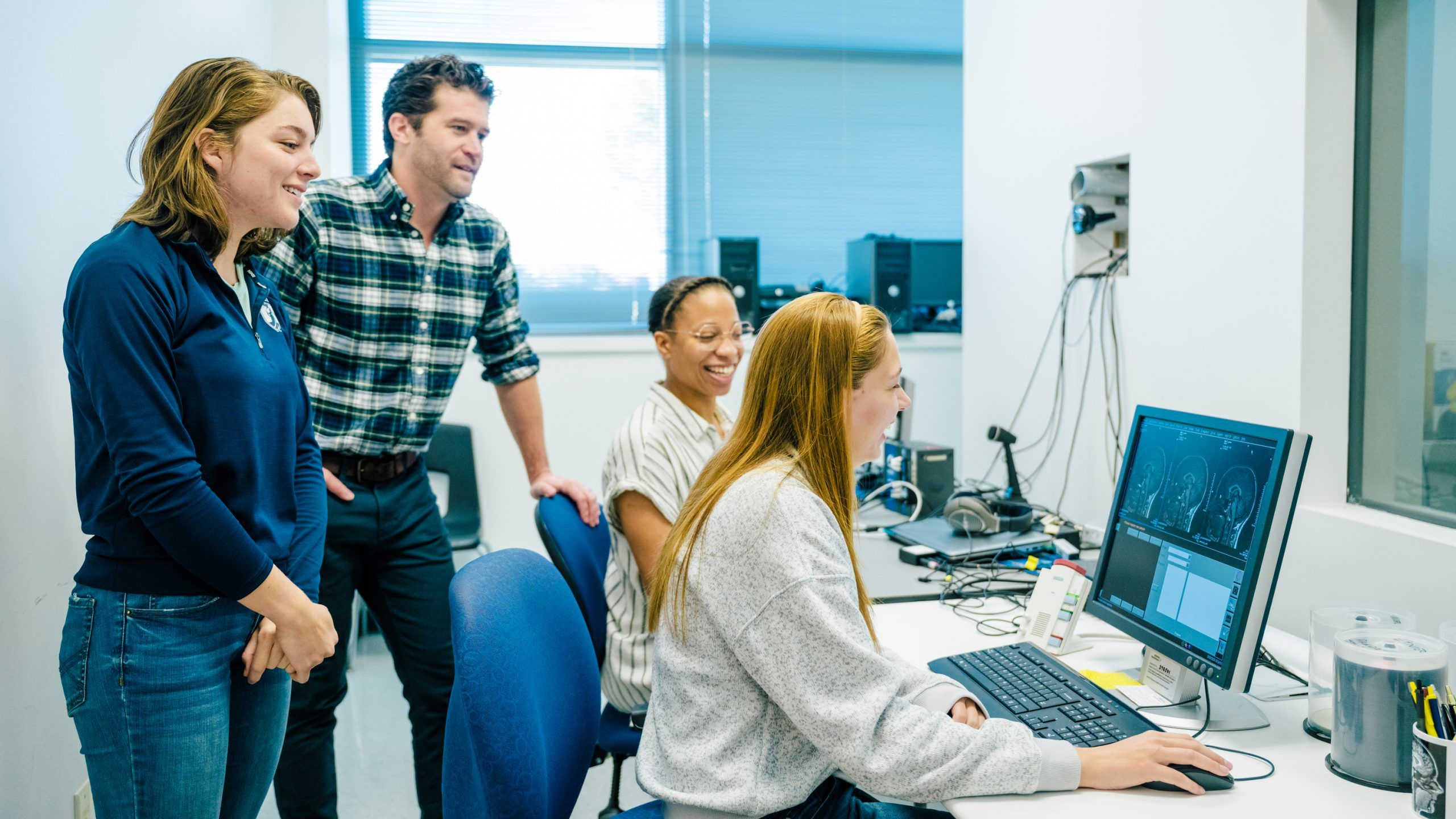
“We’re open for business. You can schedule here, you can scan here,” he says. “Top to bottom, the support is incredible.”

  • UNCG students using the MRI machine
  • Researchers reading data over the shoulder of a student
  • Students and professor reading the findings of a brain MRI
  • five people standing outside of UNCG JSSN building

By Mark Tosczak
Photography by Sean Norona & Martin W. Kane
This story is part of “Tools of the Trade,” a 2023 feature

Social Media