Tools of the Trade
Tools of the Trade
Observation has always been fundamental to research – in the field or in a tightly controlled lab.
At the Joint School of Nanoscience and Nanoengineering, or JSNN, millions of dollars’ worth of major scientific instruments are allowing faculty and students from across two universities to drive their scholarly projects forward with powerful, precise observation.
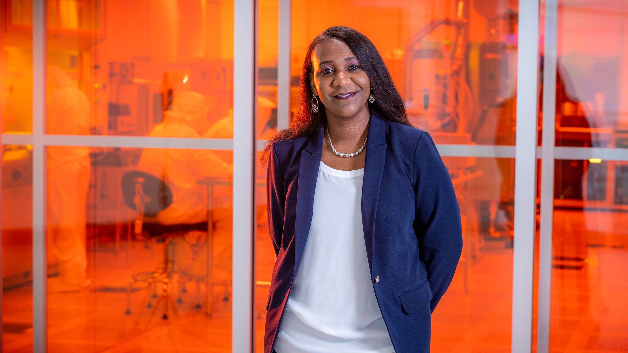
“Just for them to have access to these facilities, it enables us to move the research quicker,” says JSNN Dean Sherine Obare. It also facilitates collaborations. “Easily a fourth of the usage comes from industry partners.”
Among the major scientific instrumentation that now lives at JSNN are an MRI, a micro-CT scanner, and electron microscopes – including one that allows researchers to see objects as small as a single atom.
The multiple instruments together can give researchers a more complete view. “I need all that information together to really map out exactly what’s going on,” Dr. Obare says.
Without those instruments, researchers in kinesiology, anthropology, biology, nanoscience, and other fields would have to drive to other universities and pay hundreds of dollars per hour for access to equipment or try to negotiate access from busy hospitals.
“Even when researchers get access to those instruments, the time is restricted due to demand,” UNCG’s Vice Chancellor for Research and Engagement Terri Shelton notes. “You might only be able to get access late at night. Logistics like those can make it difficult to recruit research subjects and are disruptive for researchers and their students.”
The presence of these scientific instruments makes UNCG a more attractive destination when Obare and other leaders are recruiting new faculty and graduate students.
“The fact that we have these facilities at the Joint School opens doors for us to attract really high-quality faculty because they know that what they need is going to be in the building,” Obare says. “And faculty who are on the main campus at UNCG know they can just hop in the car and go three miles down the road – it means that they’re going to be able to easily get data.”
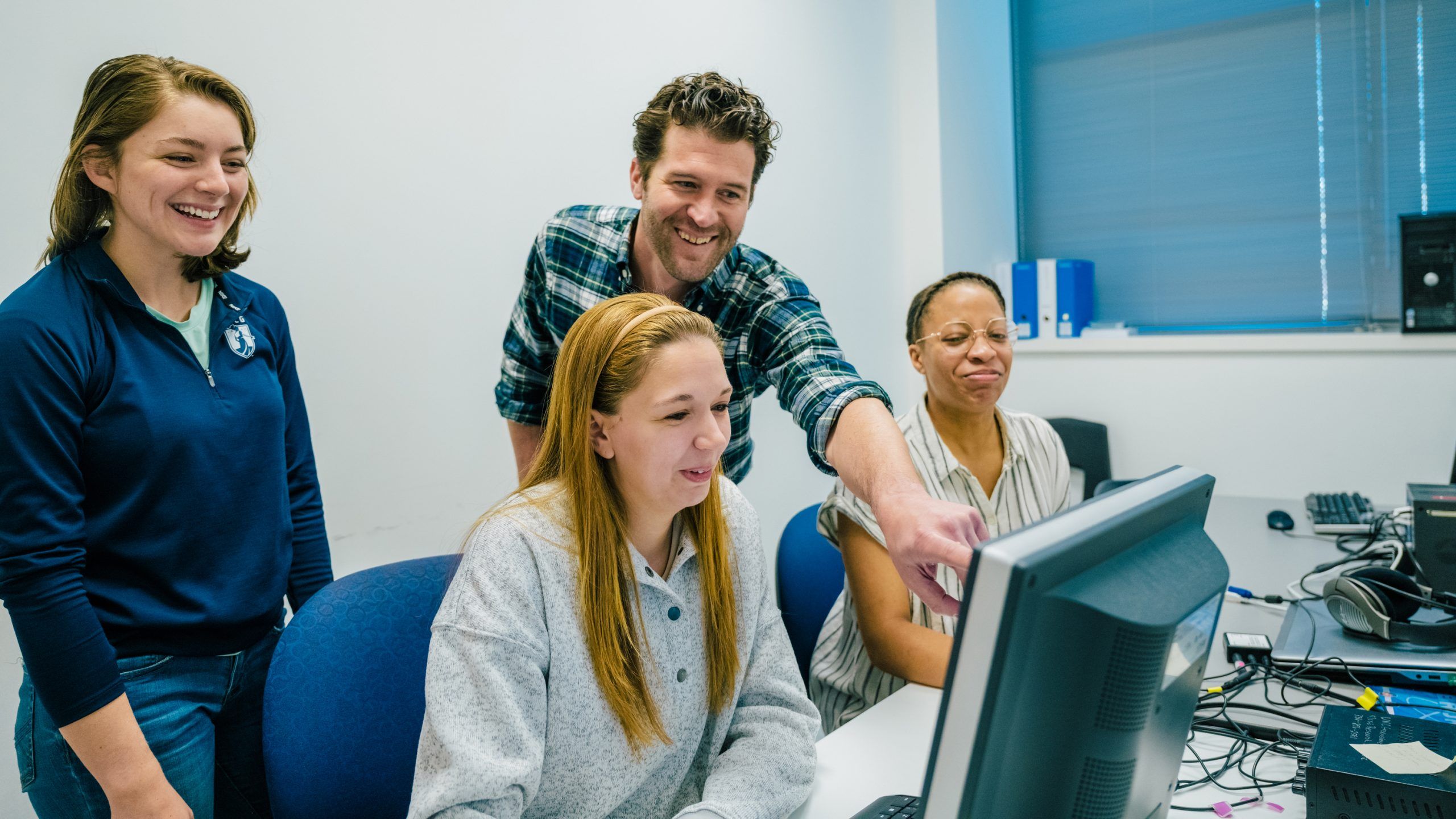
Students from UNCG and NC A&T – especially graduate students who may one day themselves be leading research labs at universities – get hands-on access to the equipment, facilitating their research and giving them the opportunity to develop expertise with the devices, she says.
“There’s a humongous gap in knowledge, in terms of student training, when a student just takes a sample and sends that sample for analysis at a different location without being physically there,” says JSNN Dean Obare, “versus them actually preparing their sample and being able to image it themselves.”
Micro-CT insights
Paleontologist and biologist explore how mammals change over time, with boost from NSF major instrumentation grant
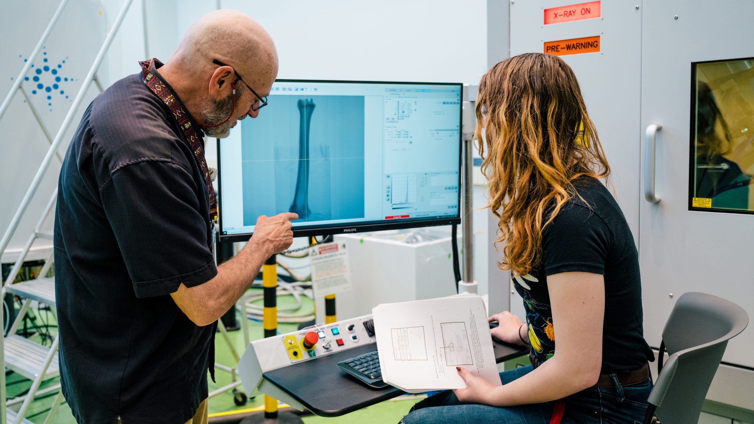
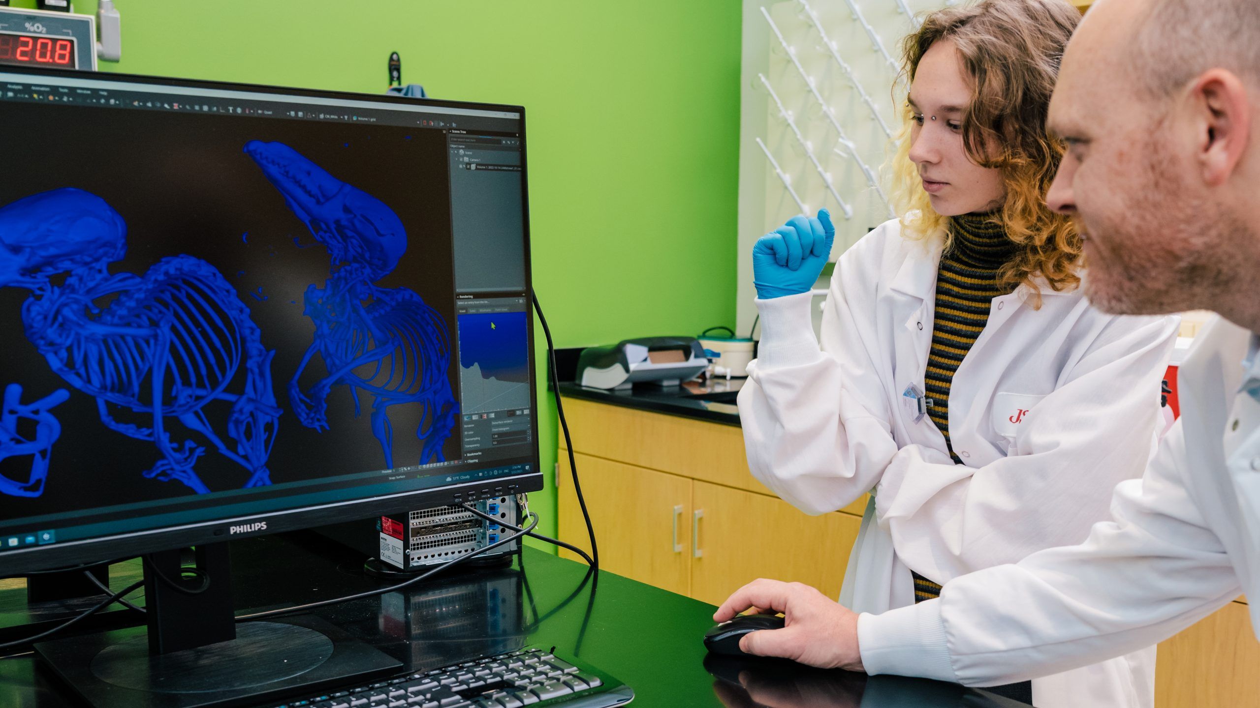
For the last few years, vertebrate paleontologist Robert Anemone would take fossils he was studying, drive more than an hour to Duke University, and pay $120 per hour for time on a micro-CT scanner.
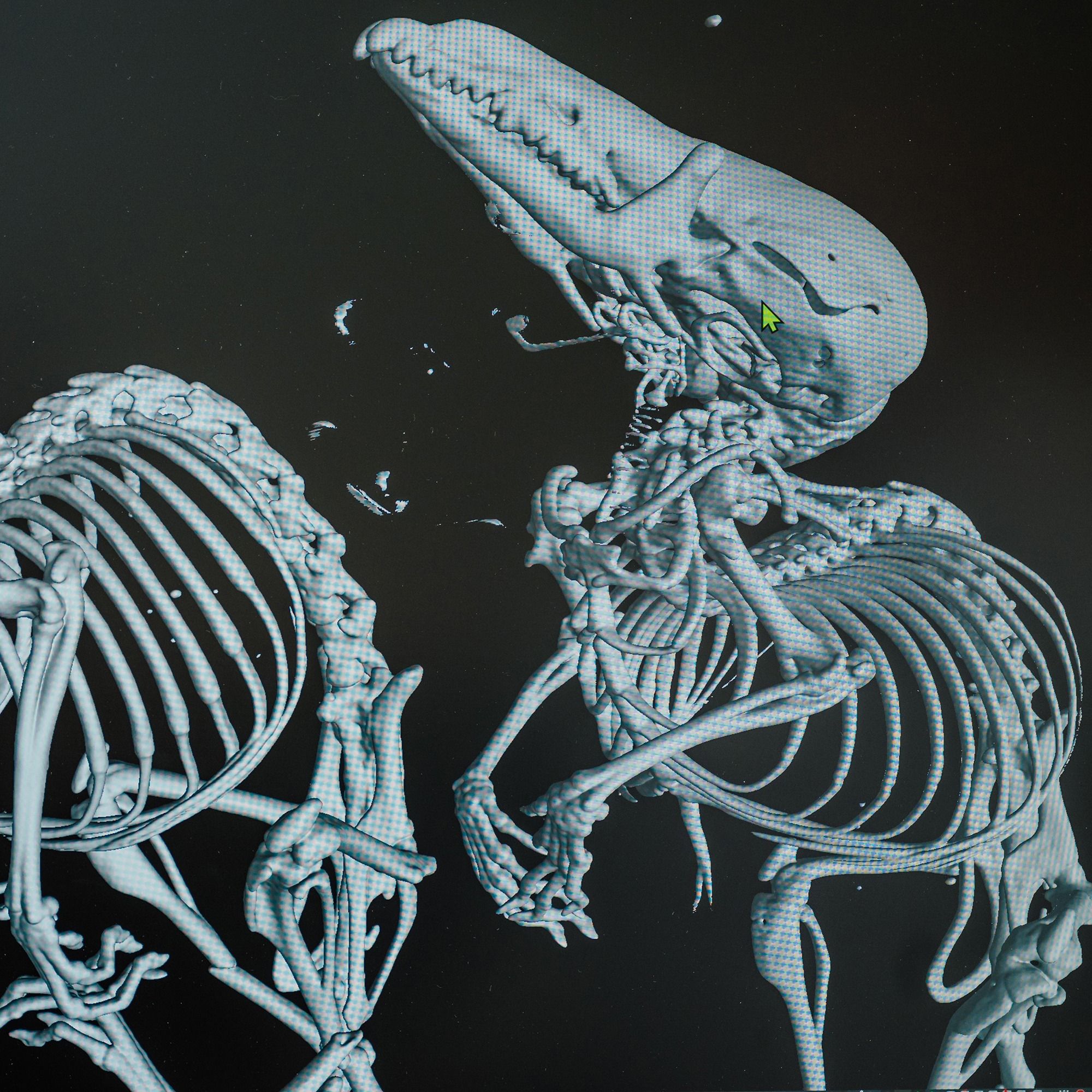
Computerized tomography scanners, long used in medicine, have become a standard research tool for paleontologists and other scientists interested in the anatomical structure of plants and animals.
“It really has revolutionized paleontology,” Dr. Anemone says.
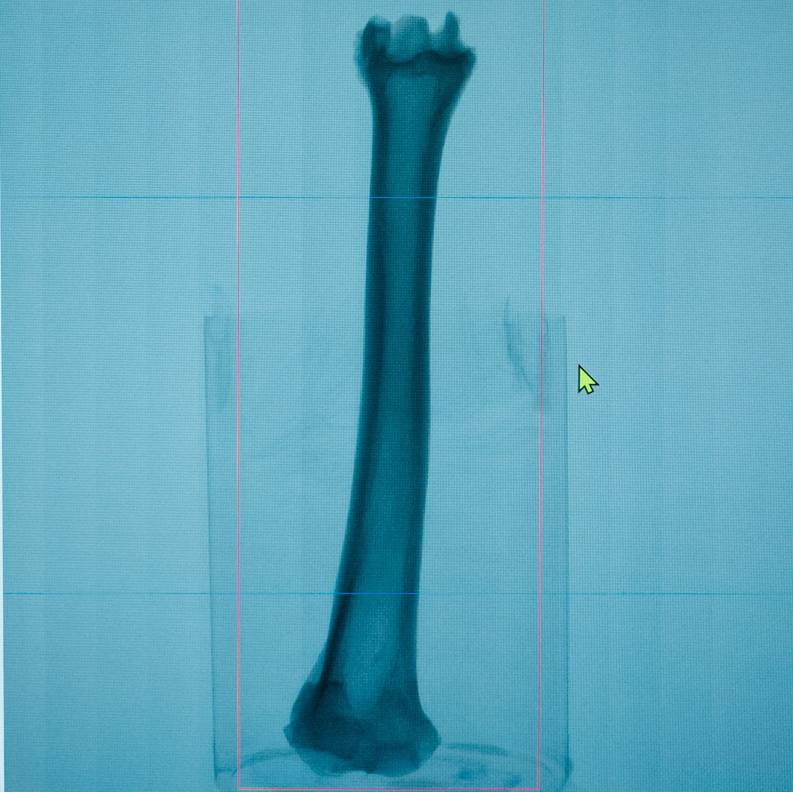
A CT scanner works by taking thousands of x-rays of a fossil or bone sample at different angles. A computer knits together those images to create a precise, three-dimensional rendering.
That enables scientists like Anemone to precisely visualize, measure, and analyze skeletal structures. Sometimes, Anemone says, he doesn’t even have to separate the fossil from the rock it’s preserved within.
But driving to Duke ate up precious time and research funds.
Biologist Bryan McLean is interested in both evolutionary changes in mammals over long time periods, as well as short-term changes in individuals from one season to another.
He’s studied how rodents around the world adapt in both similar and different ways to underground environments, including how their bodies change to allow them to dig. Some species use their forelimbs, some their hindlimbs, others make more use of their mouths.
“We’re interested in the nuance that goes along with that,” he says. “The extent to which there’s trade-offs – the forelimb becomes enlarged, the other limbs aren’t.”
By studying the animals’ anatomy, he hopes to gain insights into how the environment can shape evolution.
He’s also interested in how short-term variations, such as changes in food availability and the weather, can prompt physiological changes – specifically, ones he can track through changes in anatomy.
Into the Tissue
MRI powers studies of ACLs, Alzheimer’s and athletes – and unique educational opportunities
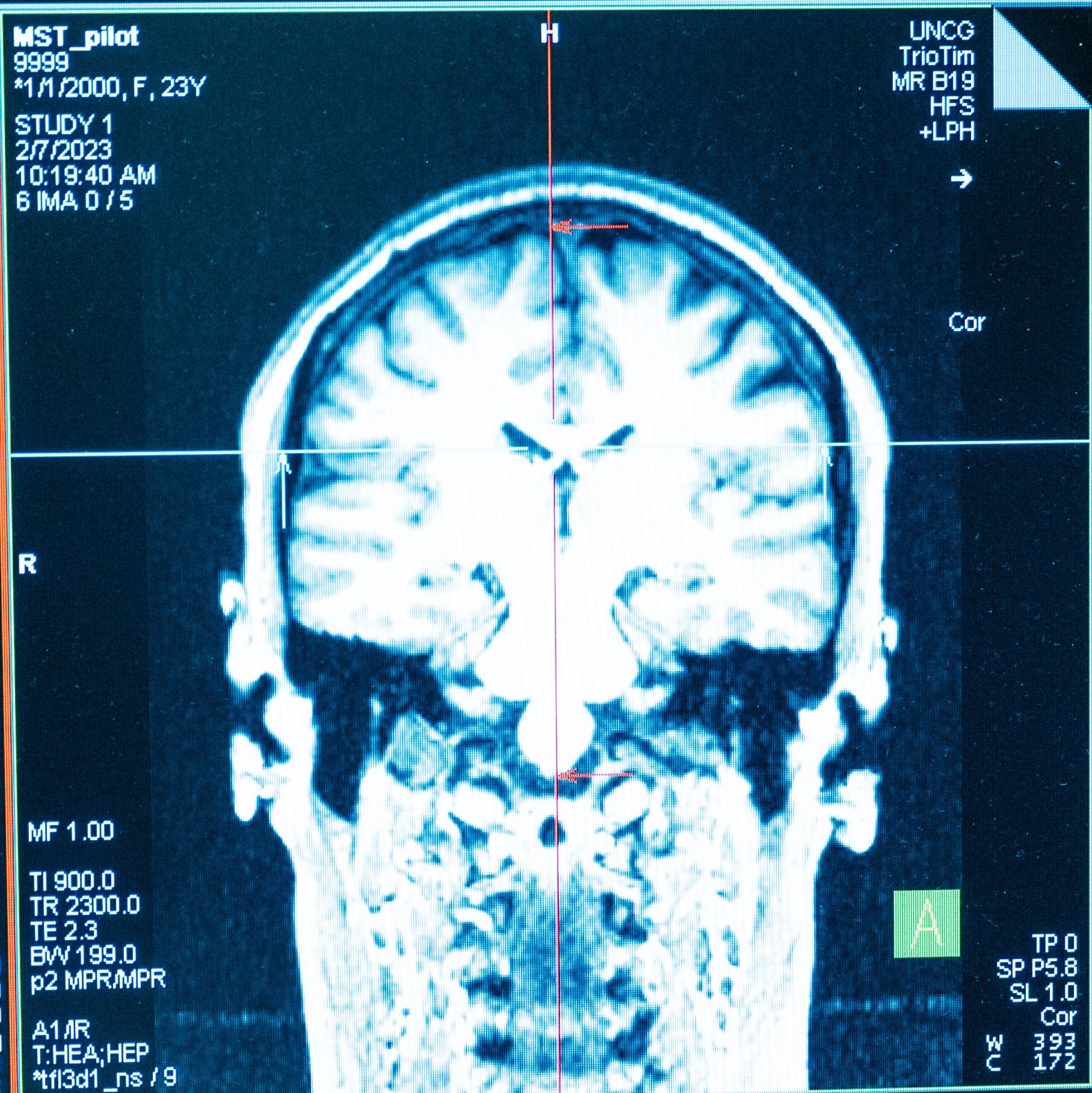
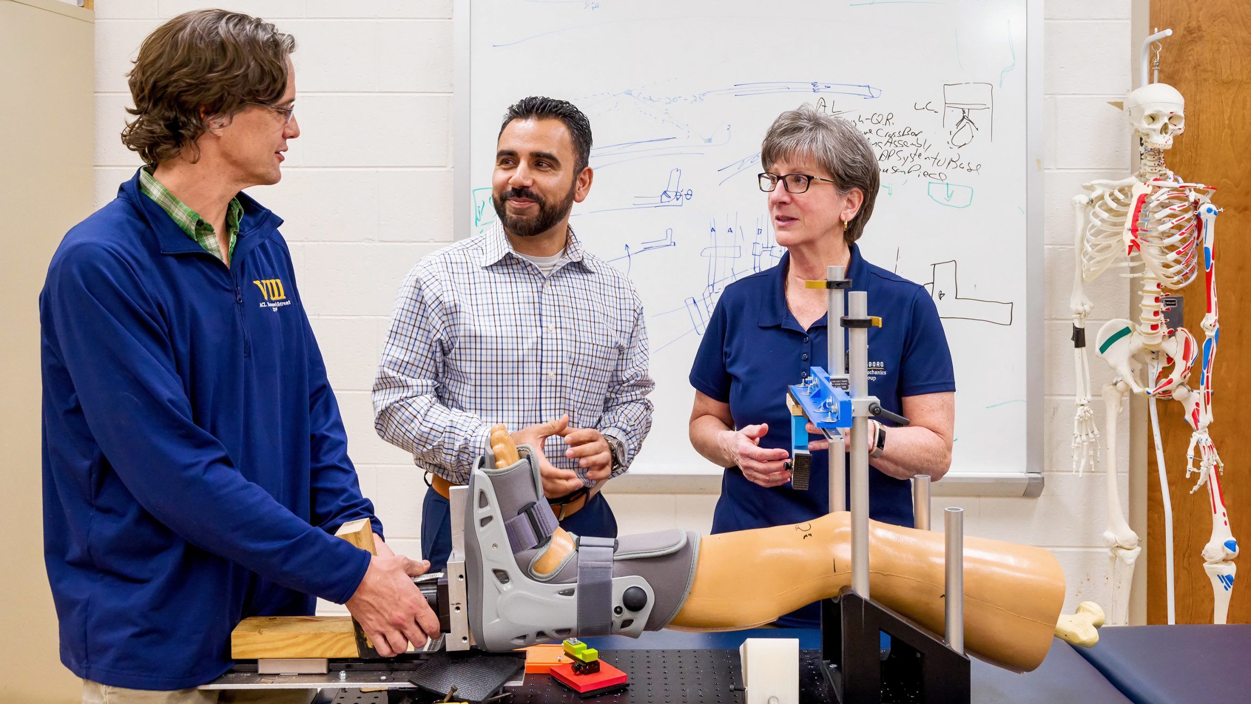
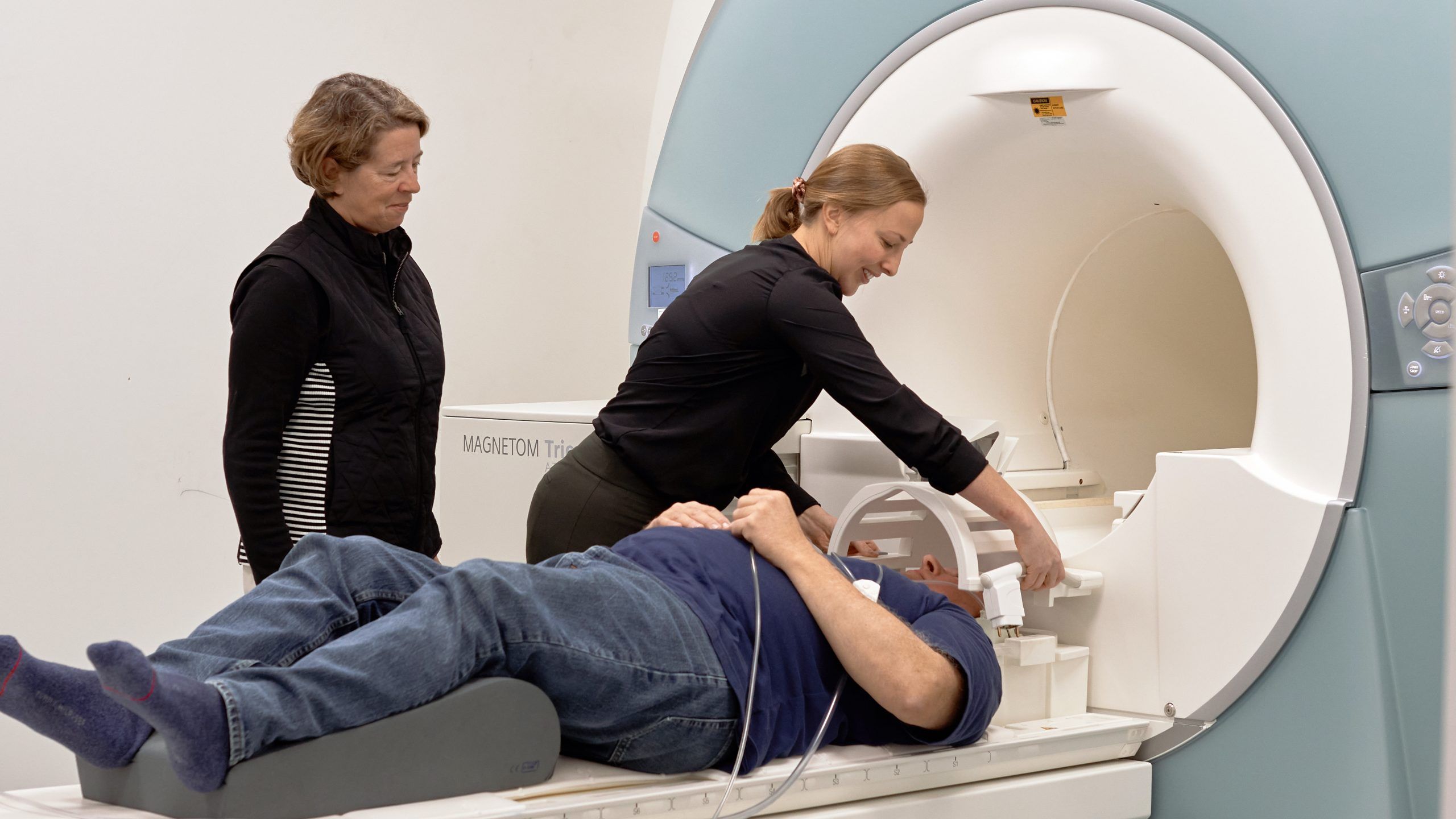
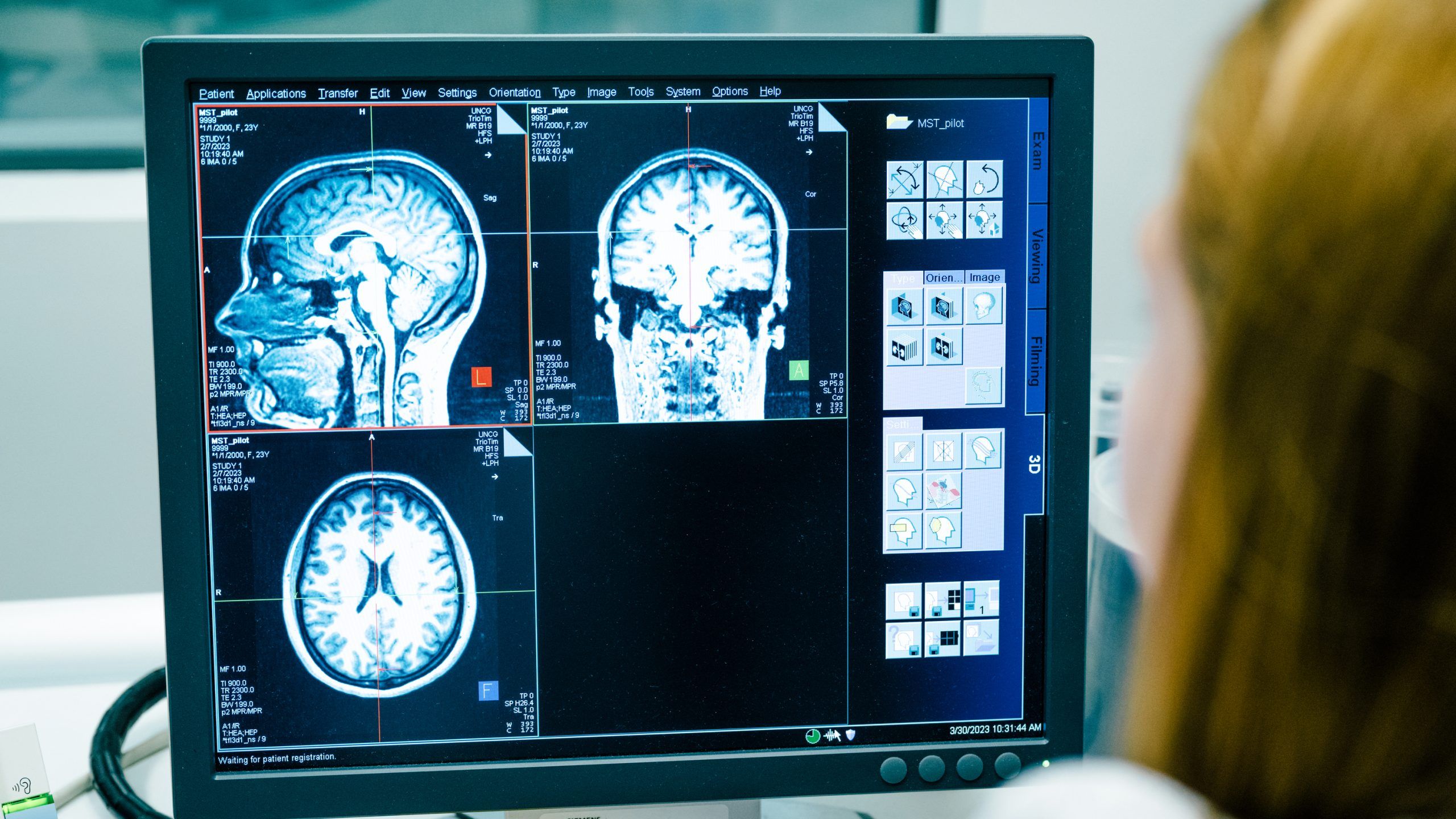
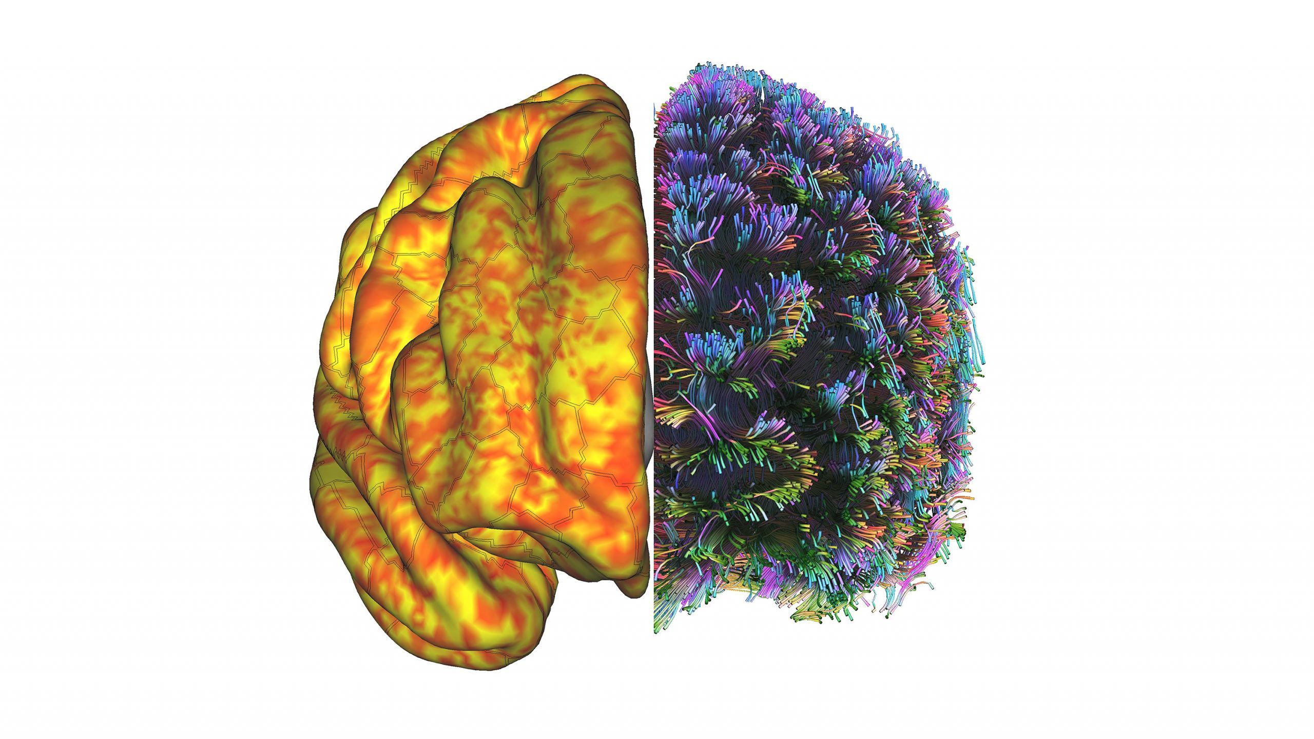
UNCG researchers are using a magnetic resonance imaging machine, or MRI, to peer inside joints and take pictures of study volunteers’ brains. Their goal is to better understand how our bodies and brains function – and why things go wrong – to find ways to prevent common problems.
“It never ceases to amaze me that here at UNCG we have access to this amazing tool to help us ask the questions that we really want to ask,” says Randy Schmitz, professor of kinesiology and director of musculoskeletal imaging at the Gateway UNCG MRI Center, where the MRI is housed.
“This has torn down barriers,” he says. “Our researchers no longer have to go to a medical institution here in town or another academic institution 60 miles away.”
MRIs create high-resolution images of tissues that have water in them – virtually all tissues in the human body.
Powerful magnets in the machine cause protons in water molecules to align, and then the machine emits radio waves that tickle those water molecules. The water molecules give off energy the machine detects to create pictures of tissues. It can even measure blood flow in the brain.
At the nanoscale
Atomic-scale imaging paves the way for sustainable nanotech, draws in graduate students
Atoms make up everything we can see – from one-celled organisms only visible under a microscope to miles-high mountains. But individual atoms and the complex structures they can form are too small to be seen by the naked eye, even under the most powerful optical microscopes.
That’s a challenge for scientists like the JSNN’s Dr. Hemali Rathnayake, who has devoted her career to studying and creating materials at the atomic scale.
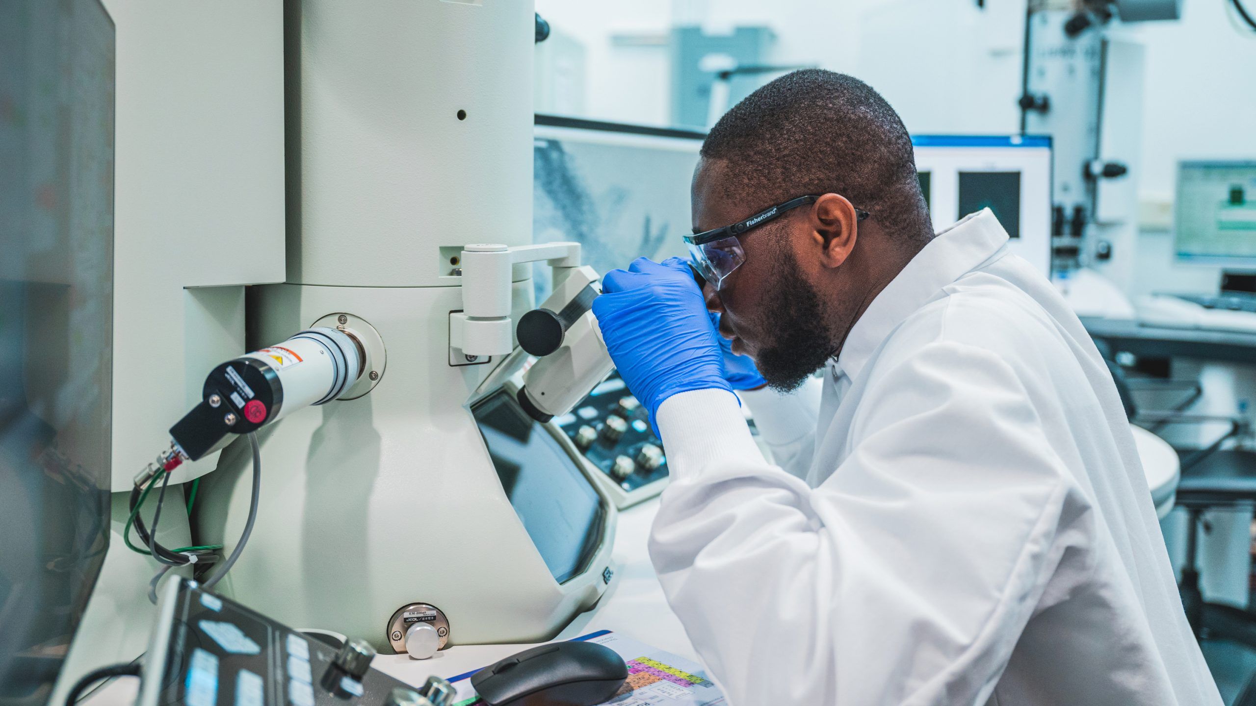
To see, for example, openings that are big enough to let through some molecules but too small to let through others, she and her research collaborators use powerful microscopes that don’t rely on light at all. Instead, they use beams of electrons.
Nanoscience is the study of objects and phenomena on the nanoscale, or one-billionth of a meter. For comparison, a sheet of paper is about 100,000 nanometers thick. Or, put another way, if the diameter of the earth was one meter, then one nanometer would be about the diameter of a marble.
“If we didn’t have access to these electron microscopes, then actually most of our research would not move forward in the nanoscale,” Rathnayake says. “We would be unable to do a lot of new science.”
